


CCL4 Liver Fibrosis Model
Test whether your compound reverses established fibrosis
Model acute liver injury, advanced fibrosis, and fibrosis reversal in a highly reproducible CCl4 liver fibrosis model, with a validated multi-endpoint readout built for antifibrotic drug screening.
A Reproducible CCl4 Model of Liver Fibrosis for Antifibrotic Discovery
Liver fibrosis is a wound-healing response to chronic injury that drives excessive extracellular matrix deposition and progresses toward cirrhosis, liver failure, and portal hypertension. The CCl4 model of liver fibrosis reproduces the inflammation, regeneration, and fiber formation seen in human disease, which makes it a dependable platform for antifibrotic drug screening.
Repeated carbon tetrachloride exposure damages hepatocytes and activates the fibrogenic cascade, laying down collagen-rich scar tissue across the liver in a predictable, time-dependent way. Because the injury follows a well-characterized course, the model gives drug developers a reliable window in which to evaluate a candidate’s effect on fibrosis progression and reversal.
Melior runs the CCl4 liver fibrosis model with a validated, multi-endpoint readout that quantifies both injury and scarring, so findings translate into real confidence in a compound’s antifibrotic activity. The result is a clinically relevant model whose features closely mirror the inflammation and scarring seen in human liver fibrosis.
Why Researchers Choose Melior's CCl4 Liver Fibrosis Model
- Highly reproducible fibrosis. IP CCl4 drives a consistent fibrotic phenotype within four weeks.
- Complete readout in one study. Enzymes, collagen, hydroxyproline, and histopathology capture injury and fibrosis.
- Validated internal benchmark. Our historical control dataset benchmarks candidates without a positive control.
- Flexible study designs. Prophylactic, intervention, and regression dosing test prevention through fibrosis reversal.
This model is 1 of more than 60 models included in our theraTRACE® platform
Ready to get started or looking for a custom model?
Contact us today for more information about our bespoke research models and to discuss how we can help you answer your unique research questions.





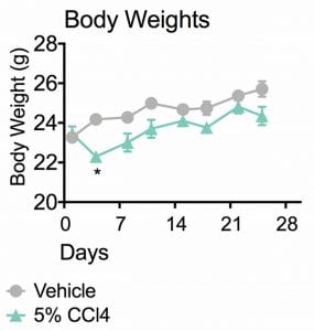
Figure 1. Body Weights. This study was conducted in male Balb/c mice (N=10). After the first two doses, animals receiving 5% CCl4 had a significant drop in body weight at day 4. Though these animals continued to grow, they remained 2-5% smaller than their vehicle counterparts. Data are mean ± SEM; *p<0.05 compared to vehicle control.
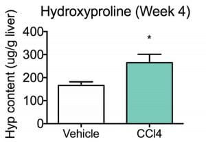
Figure 2. Hydroxyproline Content (week4). Hydroxyproline (4-hydroxyproline, Hyp) is a common non proteinogenic amino acid. Hydroxyproline in tissue hydrolysates is an indirect measure of the amount of collagen deposition. In line with the results of PSR collagen staining, hepatic Hyp content levels in CCl4 treated animals were significantly higher than vehicle treated animals. Data are mean ± SEM; *p<0.05 compared to vehicle control.
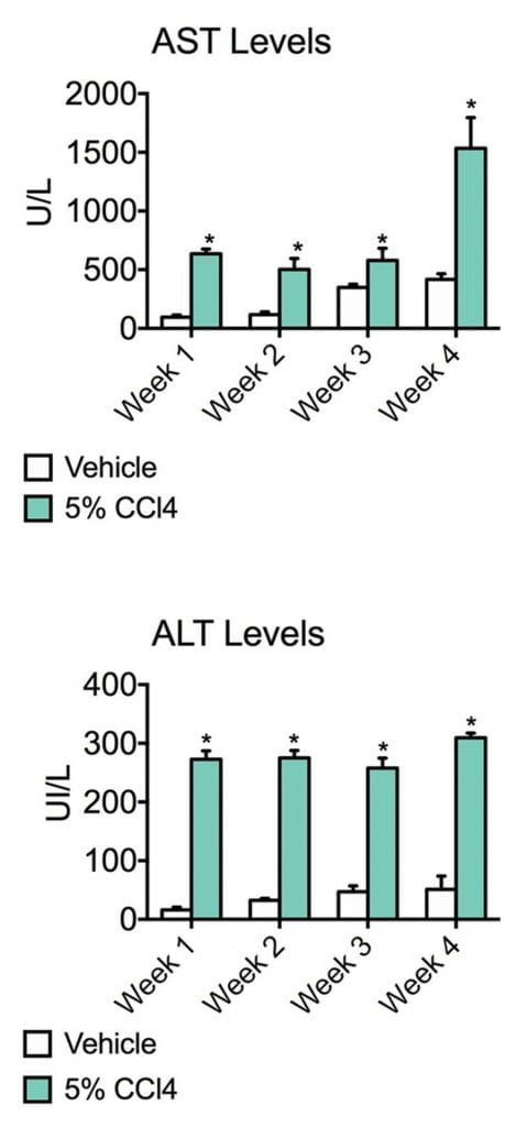
Figure 3. AST levels and ALT levels. Aspartate transaminase (AST) and alanine transaminase (ALT) are commonly measured clinical biomarkers of liver health. Both AST and ALT levels were significantly elevated in CCl4 administered animals for the entire duration of the study, suggesting that liver damage has occurred. Data are mean ± SEM; *p<0.05 compared to vehicle control.
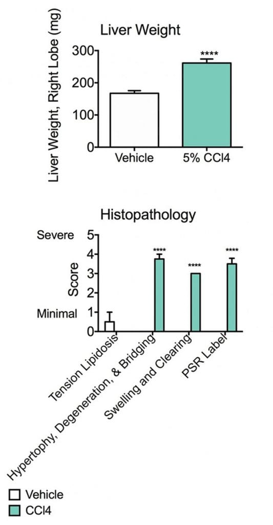
Figure 4. Liver weight and Histopathology. The right lobe of the liver was weighed prior to homogenization; livers of the CCl4 group were significantly heavier than those of vehicle animals. This may be related to hypertrophy and increased swelling found in the pathological analysis. Data are mean ± SEM; ****p<0.0001 compared to vehicle control.
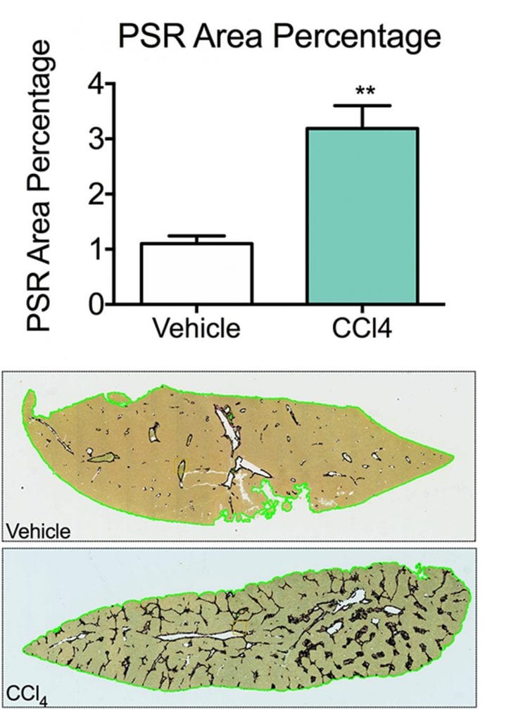
Figure 5. PSR Area Percentage and Staining. Picrosirius Red (PSR) staining is a commonly used histopathology technique to visualize collagen/fibrosis in tissue sections. The percentage of PSR detected in liver sections from animals receiving CCl4 was significantly higher than that of vehicle animals, suggesting that fibrosis has been achieved. Sections of liver were also scored for pathology (0= no finding, 1= minimal, 2= mild, 3= moderate, 4= marked, 5= severe). Data are mean ± SEM; **p<0.01 compared to vehicle control.
These data illustrate the clinically relevant presentation of liver fibrosis in this animal model. The features described above are highly reminiscent of those seen in human liver fibrosis.
Get the data you need with customized experiments and expert advice.
Frequently Asked Questions
Liver enzymes (AST, ALT) and hepatic hydroxyproline content are measured and both increase in this CCl4 mouse model. Histology, including PSR morphometry and severity scoring, is one of the most accurate ways to confirm fibrosis.
Yes. We work with histology labs that handle the full tissue processing and analysis for the CCl4 model on request.
Yes. A post-CCl4 regression arm can be added to the same study to evaluate whether a compound reverses fibrosis after injury has been established.
A standard CCl4 liver fibrosis model study runs roughly 13–16 weeks end to end: acclimation (1 week), four-week CCl4 induction, a 2–4 week treatment window, necropsy and tissue processing (1–2 weeks), histology and data analysis (2–4 weeks), and final reporting (1–2 weeks).
Citations
- Nevzorova, Y.A., Luedde, T., Zimmermann, H. et al. Liver Fibrosis—From Mechanisms of Injury to Modulation of Disease. Front. Med. 8, 814496 (2022). https://doi.org/10.3389/fmed.2021.814496